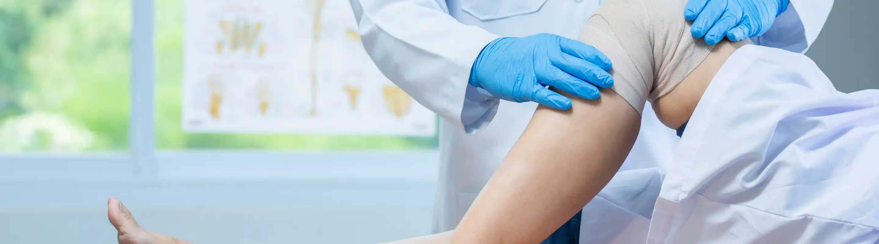
Successful Knee Pain Treatment in Pittsburgh
Knee Fracture Treatment
A knee fracture is a break in the bone of your knee. Your knee has three bones: the thighbone (femur), shinbone (tibia), and kneecap (patella), and a fracture can occur in any of these bones. In younger individuals, these fractures are caused by high-energy injuries, such as a motor vehicle accident or a sports injury. In older people, the most common cause is weak and fragile bones.
There are a few different knee fracture types, including:
- Distal femur fracture - The distal femur (top part of the knee joint) is part of the femur bone that flares out, similar to the mouth of the funnel. A distal femur fracture is a break in the thighbone that occurs just above your knee joint.
- Femoral shaft fracture - A femoral shaft fracture is a break that occurs anywhere along the femoral shaft, which is the long, straight part of the femur.
- Fractures of proximal tibia - A proximal tibial fracture is a break in the upper part of the shin bone or tibia. Proximal tibial fractures may or may not involve the knee joint, although fractures that enter the knee joint may cause joint imperfections, irregular joint surfaces, and improper alignment in the legs.
- Tibial shaft fractures - A tibial shaft fracture is a break that occurs along the length of the tibia or shin bone (larger bone of the lower leg) between the knee and ankle joints. These fractures can occur while playing sports such as soccer and skiing.
While you should visit your orthopaedic specialist to be sure if you have fractured your knee, some knee fracture symptoms you can look out for are:
- Intense pain in the knee or leg.
- Inability to put weight on the affected leg.
- Knee deformity, such as your knee appearing sunken in or out of place.
- Swelling and bruising around the knee joint.
- Knee instability, such as feeling like your knee is going to "give out".
If you think you may have fractured your knee, it's important to seek medical attention right away. Your orthopaedic specialist can diagnose a knee fracture through your medical history, a physical examination, and other diagnostic imaging tests like X-rays. X-rays are taken to know whether the bone is intact or broken, and they are helpful to know the type and location of the fracture. Your orthopaedic doctor may also recommend a computerized tomography (CT) scan to know the severity of the fracture.
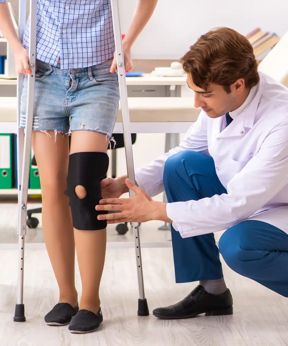
There are many treatment options for knee fractures, both non-surgical and surgical. Non-surgical treatment involves skeletal traction (placement of pin into the bone in order to realign broken bones) and the use of casts and braces. Knee fracture surgery options involve internal and external fixation procedures, such as:
- Intramedullary nailing - In this internal fixation procedure, a specially designed metal rod is placed into the canal of the femur. Then the nail is passed on to reach the fracture site and keep it in place. The rod is secured in place with screws at both ends.
- Plates and Screws fixation - In this internal fixation procedure, your surgeon will reposition the broken bone ends into a normal position and then uses special screws or metal plates on the outer surface of the bone to hold the bone fragments in place.
- External fixation - During this procedure, metal pins or screws are inserted into the middle of the femur and tibia and are attached to a device outside the skin to hold bone fragments in place to allow alignment and healing. If your bone is fractured in many pieces, a plate or rod is fixed at both ends of the fracture to maintain the overall shape and length of the bone in place while it heals.
If you have fractured your knee, seek medical assistance as soon as possible, as early intervention is the best way to ensure a successful recovery.
Dr. Brian Kelly, an experienced orthopaedic surgeon, is an excellent resource for any knee pain you may be experiencing. Reach out Brian J. Kelly MD for comprehensive care and effective injury treatment.
Acclaimed Knee orthopaedic Surgeon In Pittsburgh
Patellar Dislocations
The patella, also known as the kneecap, is a small bone in the front of the knee that protects the knee joint. The patella sits in a groove at the end of the femur (thighbone) and glides up and down as you bend and straighten your knee. If the kneecap partially comes out of the groove, it is called subluxation; if the kneecap completely comes out, it is called dislocation.
Patellar dislocation occurs when the patella (kneecap) pops out of the groove completely and slides to the side. This misalignment can damage the underlying soft structures, such as muscles and ligaments, that hold the kneecap in place. Once damaged, these soft structures are unable to keep the patella in position.
Patellar dislocations are more common in young athletes, and particularly affect women because the wider pelvis creates a lateral pull on the patella. Some of the causes of patellar dislocation include a direct blow or trauma to the knee, twisting of the knee while changing direction, muscle contraction, and congenital defects.
Symptoms of patellar dislocation can include:
- Knee pain and tenderness.
- A popping or snapping sensation at the time of injury.
- Swelling around the knee joint.
- Knee instability.
- Inability to straighten the leg fully.
- Discoloration of the injured area.
- Numbness below the knee.
If you think you have injured your knee cap, it is important to see an orthopaedic specialist for an evaluation. Your orthopaedic doctor will examine your knee and suggest diagnostic tests such as an X-ray, CT scan, or MRI scan to confirm the condition and provide treatment.
There are non-surgical and surgical ways of treating patellar dislocation. Your specialist will likely recommend non-surgical treatment options before proposing surgery. This can include conservative treatments like:
- PRICE (protection, rest, ice, compression, and elevation).
- Non-steroidal anti-inflammatory drugs and analgesics to treat pain and swelling.
- Braces or casts that will immobilize the knee and allow the MPF ligament to heal.
- Footwear to control gait while walking or running and decrease the pressure on the kneecap.
If patellar dislocation does not respond to conservative treatments and becomes a recurrent problem, your orthopaedic surgeon may recommend surgical treatments, such as:
- Lateral release - In this procedure, your surgeon will loosen or release the tight lateral ligaments that pull the kneecap from its groove, increase pressure on the cartilage and cause dislocation.
- Medial patellofemoral ligament reconstruction - This procedure involves the torn MPF ligament being removed and reconstructed by using a graft. Grafts are usually harvested from the hamstring tendons, located at the back of the knee, and are fixed to the patella tendon using screws. The grafts are either taken from your own body (autograft) or a donor (allograft).
- Tibia tubercle realignment or transfer - The tibial tubercle is a bony attachment below the patella tendon, which sits on the tibia. In this procedure, the tibial tubercle is moved towards the center and held by two screws. The screws hold the bone in place so that it can heal faster and prevent the patella from sliding out of the groove.
These procedures are highly effective and can be performed using an arthroscope. Dr. Brian Kelly and his team are experienced in providing each of these procedures to relieve pain and restore mobility. If you have injured your kneecap, put your trust in the top orthopaedic knee specialist near you at the office of Brian J. Kelly MD.
Frequently Asked Questions
How can I prevent patellar dislocation?
The best way to prevent patellar dislocation is to warm up properly before playing sports or exercising. Wearing the proper shoes for your sport and ensuring that they fit well can also help. If you have had a previous patellar dislocation, your orthopaedic surgeon may recommend wearing a knee brace during activities.
What does post-operative care for knee fracture surgery entail?
After knee surgery, your orthopaedic surgeon may suggest you use crutches for a few weeks, prescribe medications to control pain and swelling, and recommend physical therapy to help you return to your sports activities at the earliest. Physical therapy is recommended to control pain and swelling, prevent the formation of scar of soft tissue, and encourage collagen formation.
What is the recovery time for patellar dislocation surgery?
Recovery times will vary depending on the type of procedure performed. For example, medial patellofemoral ligament reconstruction has a longer recovery time than lateral release because it is a more complex surgery. Most people can return to work and driving within a week after medial patellofemoral ligament reconstruction. Recovery times for other procedures are typically shorter.
It is important to follow your surgeon’s instructions for a safe and successful recovery. This will usually involve physical therapy to regain range of motion and strength in the knee joint. A return to sports and other activities will be based on your progress in physical therapy and clearance from your surgeon.
How do I get started with an orthopaedic knee specialist?
Dr. Brian Kelly and his team are committed to providing you with the best possible care to help you recover from knee injuries. Dr. Kelly will work with you to create a customized treatment plan that fits your needs and lifestyle.
Don’t let knee pain interrupt your daily life, schedule an appointment today to relieve your symptoms. Contacts us by phone at 412-262-7800, or visit our office at 725 Cherrington Pkwy, Suite 200, Moon Township, PA 15208.
Knee Pain? Call Dr. Brian Kelly Today!
Don’t let knee pain keep you down. Visit the best orthopaedic surgeon near you.